
Lameness
Lameness can range from a subtle, performance limiting problem or very obvious, severe lameness.
Most forelimb lamenesses will be caused by a problem below the knee level; elbow and shoulder problems are rare.
The causes of hindlimb lameness are more varied and investigation of these cases can be more time-consuming.
Usually, the first examination of lameness is at your yard. In cases of severe lameness, we will usually only need to see a few steps of movement from the horse; however, in subtle lamenesses, we will need to see the horse walked and trotted, perform flexion tests, lunge them and sometimes even see them ridden.
If you do not have suitable facilities at home, we have all the facilities required for full lameness investigation including ridden exercise on our purpose built menage.
The benefit of clinic-based lameness investigations is that all the equipment is to hand, rather than requiring repeated trips to your yard to investigate, nerve block, x-ray or ultrasound scan your horse. This should hopefully provide a quicker route to diagnosis and treatment.
Lameness investigation
Isolating the problem
Once the lame leg is established and if there are no obvious symptoms, we will usually require nerve blocks to establish the area causing lameness.
Nerve blocks are small injections of local anaesthetic round nerves in the leg.
After the injections, the lameness examination is repeated and if the horse has improved sufficiently, we can be sure the lameness originates from the area that has been ‘numbed out’ by local anaesthetic.
Imaging
Once we have established the area causing lameness, we can then image the area to provide a diagnosis, a treatment plan and a prognosis.
Usually, radiographs (x-rays) are taken to assess the bony structures in the area.
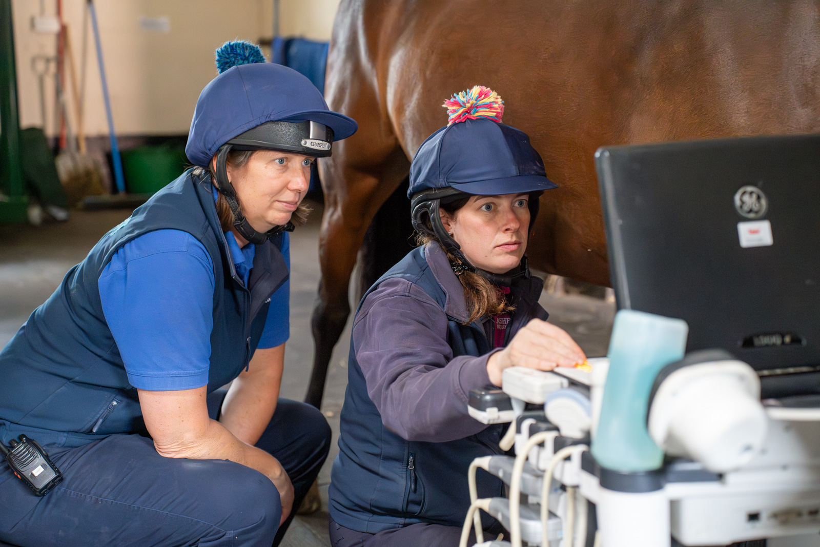
Ultrasound scanning enables us to assess for tendon or ligament damage or even damage to the surface of bones.
MRI
We have a standing Magnetic Resonance Imaging (MRI) unit.
This can be used to provide vital information about the small tendon and ligament structures of the lower limb, particularly those structures hidden in the hard hoof capsule.
Isolating the problem
Once the lame leg is established and if there are no obvious symptoms, we will usually require nerve blocks to establish the area causing lameness.
Nerve blocks are small injections of local anaesthetic round nerves in the leg.
After the injections, the lameness examination is repeated and if the horse has improved sufficiently, we can be sure the lameness originates from the area that has been ‘numbed out’ by local anaesthetic.
Imaging
Once we have established the area causing lameness, we can then image the area to provide a diagnosis, a treatment plan and a prognosis.
Usually, radiographs (x-rays) are taken to assess the bony structures in the area.
Ultrasound scanning enables us to assess for tendon or ligament damage or even damage to the surface of bones.
MRI
We have a standing Magnetic Resonance Imaging (MRI) unit.
This can be used to provide vital information about the small tendon and ligament structures of the lower limb, particularly those structures hidden in the hard hoof capsule.
Treatment plan
Nerve and joint blocks, together with imaging results allows us to formulate a treatment plan, which can be medical or surgical.
Medical treatment of lameness involves many different treatments and often we use a combination of these to provide the best result for you and your horse.
Lameness problems that require surgical intervention can be operated on in our surgical suite.
Lameness treatment options
The options for medical treatment of lameness are varied, ranging from basic through to costly options reserved for those cases not responding to conventional treatments.
We will also consider the whole horse and as such, can recommend remedial farriery, manipulative therapy and joint supplements as required.
We will always try to incorporate your wishes, your horses’ requirements, competition rules and any financial constraints into the treatment plan.
Non-steroidal anti-inflammatory drugs
Oral anti-inflammatory medications may seem old fashioned, but they are immensely beneficial to reduce swelling and pain while a simple injury heals.
They may also be used as a stand-alone treatment or until other treatments have a chance to improve the lameness.
Conventional joint medications
If a lameness has been isolated to a specific joint, we will often inject medications directly into that joint. This form of treatment can be very effective within a short period of time.
Corticosteroid medication
We have two different types of corticosteroid medication that are commonly used in joint medication, depending on which joint is affected and any required competition withdrawal periods.
We will usually see a dramatic improvement in lameness within 10 to 14 days, allowing the horse to quickly return to his normal job.
Bisphosphonates
These drugs, given as intravenous infusions, act on the damaged bone at the surfaces of arthritic joints by reducing the bone resorption associated with arthritis.
Extracorporeal shockwave therapy
This treatment uses pulsed waves of energy focussed over the area of damaged tissue reported to stimulate healing and also provide some degree of pain relief.
Biological treatments
We are also able to offer all the latest biological therapeutics available for lameness treatment.
These treatments include stem cell-based products and anti-inflammatory products, injected directly into soft tissue structures or joints.
Standing MRI scans
We’re pleased to be able to offer MRI investigation of lameness problems.
When a horse is admitted for MRI, we allow them a night of hospitalisation to settle into our hospital environment.A more settled horse means we will obtain higher quality images with less sedation.
On the day of the MRI, the horse has an intravenous catheter placed to allow sedation to be given easily.
The horse’s shoes are removed and radiographs taken to ensure no metal fragments remain in the hoof, as metal will render the images as non-diagnostic.
After this preparation, the horse enters the MRI suite. The scans are taken with the horse standing and the affected limb positioned in a large U-shaped magnet.
The images are obtained over a one to three hour period, depending on which area is to be scanned, after which they are sent electronically to an image interpreting specialist.
We usually expect the report from the consultant to be ready within 72 hours, at which point your vet will then be able to discuss the results and formulate the most appropriate treatment plan with you.
What to expect:
If your horse has been booked in for an MRI scan, We ask that you bring your horse into the hospital prior to scanning, usually the day before, to allow your horse to settle in.
This allows the horse to become accustomed to our hospital environment and means we can use lower doses of sedation. Less sedation provides us with less movement blur and better quality images.
Please bring your passport, so that we can check your horse’s vaccination status and that Section IX (exclusion from the food chain) has been signed.
We have all the feedstuffs your horse will require; however, if your horse has any allergies, please bring your own food as appropriate.
Please bring rugs and any medications or supplements that you would like your horse to have during their stay.
To facilitate repeat sedation doses, a catheter will be placed into the vein. It is standard practice to clip and sterilely prepare the area prior to catheter insertion. If you have concerns regarding this, please discuss it with a member of our team.
The appropriate shoes will need to be removed before scanning, i.e for forelimb scans both front shoes will be removed and for hindlimb scans both hind shoes will be removed.
After shoe removal, radiographs will be taken to ensure there are no small pieces of metal remaining in the foot; if there is, we will remove these prior to scanning.
The scan time will be determined by the areas to be imaged and the demeanour of your horse in our scanning environment.
After scanning, your horse will be taken back to a stable, monitored post sedation and fed when appropriate.
Once the scan is completed, the images are sent to our selected specialist for interpretation, who will endeavour to report their findings to us within 72 hours.
If your horse returns home on the same day, please ensure water is readily available and soft wet feedstuffs are given, as prolonged sedation can result in decreased bowel motility and increased colic risk.
These precautions minimise the colic risk; however, we advise monitoring your horse closely for the following 12 to 24 hours, especially with regard to faeces passed.


